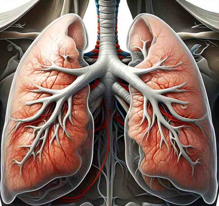
PLEURA
The pleura is a serous membrane that encloses the lungs and lines the thoracic cavity. It facilitates frictionless lung movement during respiration.
LAYERS OF THE PLEURA:
The pleura has two layers:
A. Parietal Pleura (Outer Layer)
• Lines the inner surface of the thoracic wall and surrounding structures.
• Divided into 4 parts:
- Costal pleura – Lines inner ribs and intercostal muscles.
- Diaphragmatic pleura – Covers the superior diaphragm.
- Mediastinal pleura – Covers the mediastinum.
- Cervical pleura (pleural cupula) – Extends above the first rib.
B. Visceral Pleura (Inner Layer)
• Firmly adheres to the lungs, covering their surface.
• Continuous with the parietal pleura at the hilum.
C. Pleural Cavity
• A potential space between parietal and visceral pleura.
• Contains pleural fluid for lubrication.
PLEURAL RECESSES
Potential spaces for fluid accumulation in pleura are called pleural recesses.
• Costo-diaphragmatic Recess – Largest recess, between the costal and diaphragmatic pleura. This is the site for pleural fluid aspiration (thoracocentesis).
• Costo-mediastinal Recess – Near the heart, between costal and mediastinal pleura.BLOOD SUPPLY
(a) Parietal pleura:
• Arterial: Intercostal & internal thoracic arteries.
• Venous: Azygous system.(b) Visceral pleura:
• Supplied by bronchial arteries.
NERVE SUPPLY
• Parietal pleura:
- Somatic innervation (pain-sensitive).
- Intercostal nerves (costal pleura).
- Phrenic nerve (diaphragmatic & mediastinal pleura).
• Visceral pleura:
- Autonomic innervation (no pain sensation).
- Vagus & sympathetic nerves.
CLINICAL ASPECTS
-
Pleuritis (Pleurisy)
• Inflammation of pleura → Sharp pain with respiration.
• Cause: Infection, autoimmune diseases.
• Sign: Pleural friction rub on auscultation.
-
Pleural Effusion
• Excess fluid in pleural cavity → Compresses lungs.
• Types:
1. Transudate (heart failure, nephrotic syndrome).
2. Exudate (infection, malignancy).
: Diagnosis: Thoracocentesis (pleural tap).
- Pneumothorax (Collapsed Lung)
• Air in pleural cavity → Lung collapse.
• Types:
- Traumatic (rib fractures, penetrating injury).
- Spontaneous (tall, young males, smokers).
- Tension Pneumothorax – Air trapped without escape, compressing mediastinum (emergency).
• Treatment: Chest tube insertion.
-
Hemothorax
• Blood in pleural cavity, usually due to trauma.
-
Chylothorax
• Lymphatic fluid in pleural cavity, due to thoracic duct injury.