ANATOMY OF SEMINAL VESICLES
The seminal vesicles are essential components of the male reproductive system, contributing to the formation of semen.
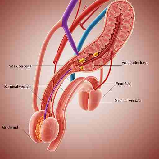
LOCATION & STRUCTURE:
- The seminal vesicles are two small, paired glands located behind the bladder and above the prostate.
- They are situated on either side of the prostate gland and are about 5-7 cm long.
- These glands are tubular, with a highly convoluted structure, and are responsible for producing a significant portion of the seminal fluid.
DIMENSIONS:
- The length of each seminal vesicle is approximately 5-7 cm, while its diameter is about 1-1.5 cm.
- The duct of each seminal vesicle joins the ductus deferens to form the ejaculatory duct.
FUNCTIONS:
- The primary function of the seminal vesicles is to produce seminal fluid, which makes up about 60-70% of the total volume of semen.
- The fluid produced by the seminal vesicles contains fructose, which provides energy for the sperm, as well as prostaglandins and proteins that aid in sperm motility and the fertilization process.
CLINICAL RELEVANCE:
- Seminal vesiculitis (inflammation of the seminal vesicles) can cause pain during ejaculation, discomfort in the pelvic region, and fertility issues.
- Seminal vesicle cysts may be asymptomatic but can lead to infertility or urinary problems if large.
- Seminal vesicle abscesses can result from infection, potentially leading to pain, fever, and infertility.
- Surgical intervention may be required in cases of cysts or abscesses, and a transrectal ultrasound is often used for diagnosis.
MICROSCOPIC STRUCTURE:
- The seminal vesicles are lined by a pseudostratified columnar epithelium, which secretes the seminal fluid.
- The secretory cells of the vesicle are richly vascularized, helping in the production and secretion of seminal fluid.