Your Account
2024 © D'vakaso.
Designed by Zeptt Technologies
Designed by Zeptt Technologies
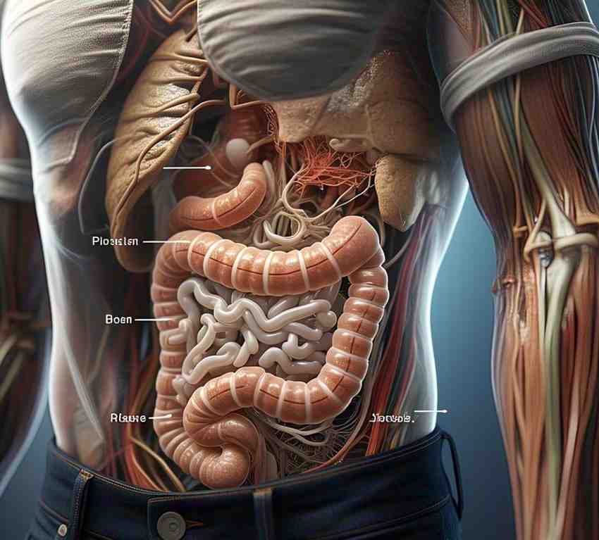
• The small intestine is the longest part of the digestive tract.
• ~6 meters in adults.
• Function:
o Digestion of food (enzymes from pancreas & bile from liver).
o Absorption of nutrients, water, and electrolytes.
DIVISIONS OF THE SMALL INTESTINE
The small intestine is divided into 3 parts:
• DUODENUM
o 25 cm / 10 inches long.
o C-shaped, retroperitoneal (except the first part).
o Receives:
Bile from the liver (via common bile duct).
Pancreatic enzymes (via the pancreatic duct).
o Divided into 4 parts:
(a) Superior (First) part – Connected to stomach.
(b) Descending (Second) part – Has major duodenal papilla (entry for bile & pancreatic juices).
(c) Horizontal (Third) part – Crosses the aorta & IVC.
(d) Ascending (Fourth) part – Joins the jejunum at the duodenojejunal flexure.
• JEJUNUM
o ~2.5 meters / 8 feet long.
o Located in the left upper quadrant (LUQ).
o Thicker wall, more vascular, and has numerous circular folds (plicae circulares) for absorption.
• ILEUM
o ~3.5 meters / 11 feet long.
o Located in the right lower quadrant (RLQ).
o Thinner wall, fewer folds, contains Peyer’s patches (lymphoid tissue).
o Ends at the ileocecal junction (joins large intestine).
STRUCTURAL MODIFICATIONS FOR ABSORPTION
The small intestine has specialized features that increase surface area for absorption:
• Plicae circulares – Permanent circular folds that slow down food movement.
• Villi – Finger-like projections lined with absorptive cells.
• Microvilli – Tiny projections on villi, contain enzymes for digestion.
BLOOD SUPPLY & VENOUS DRAINAGE
• Arterial supply: Superior mesenteric artery (SMA).
• Venous drainage: Superior mesenteric vein (SMV) → Portal vein.
LYMPHATIC DRAINAGE
• Lacteals in villi absorb dietary fats → Transported via lymphatic system.
• Lymph drains into superior mesenteric lymph nodes → Thoracic duct.
NERVE SUPPLY
• Sympathetic (Inhibitory) – From splanchnic nerves & celiac plexus.
• Parasympathetic (Stimulatory) – From vagus nerve (CN X).
CLINICAL CORRELATIONS
• Duodenal Ulcers
o Common in the 1st part of the duodenum due to H. pylori infection & acid exposure.
• Malabsorption Syndromes
o Celiac disease – Autoimmune reaction to gluten, damages villi.
o Lactose intolerance – Deficiency of lactase enzyme leads to bloating & diarrhea.
• Small Bowel Obstruction
o Caused by adhesions, hernias, or tumors → Leads to vomiting & abdominal pain.
• Crohn’s Disease
o Chronic inflammation, commonly affecting the ileum, leading to diarrhoea & weight loss.