Your Account
2024 © D'vakaso.
Designed by Zeptt Technologies
Designed by Zeptt Technologies
Hearing is the physiological process of perceiving sound through the auditory system. It involves the transmission and transduction of sound waves into electrical signals, which are interpreted by the auditory cortex of the brain. In Ayurveda, hearing is associated with the Shabda Indriya, which is a function of Akasha Mahabhuta and is governed by Vata Dosha, particularly Prana Vata and Vyana Vata.
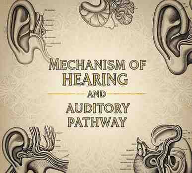
ANATOMY OF THE EAR
The ear is divided into three major parts:
External Ear – auricle (pinna) and external auditory canal
Middle Ear – tympanic membrane and auditory ossicles (malleus, incus, stapes)
Inner Ear – cochlea, vestibule, and semicircular canals
MECHANISM OF HEARING
EXTERNAL EAR FUNCTION
Collects and directs sound waves into the external auditory canal.
Sound waves strike the tympanic membrane and cause it to vibrate.
MIDDLE EAR FUNCTION
Vibrations from the tympanic membrane are transmitted to the ossicles.
The ossicles amplify the vibrations and transmit them to the oval window of the cochlea.
The tensor tympani and stapedius muscles protect the ear from loud sounds by dampening movement of ossicles (acoustic reflex).
INNER EAR FUNCTION (COCHLEA)
The cochlea contains perilymph (in scala vestibuli and scala tympani) and endolymph (in scala media).
Vibrations transmitted to the oval window create pressure waves in perilymph.
These waves cause displacement of Basilar membrane where hair cells of Organ of Corti are located.
The tectorial membrane contacts hair cells, leading to their deflection.
Deflection opens mechanically gated ion channels → leads to depolarization of hair cells.
Neurotransmitter (glutamate) release stimulates afferent neurons of cochlear nerve.
AYURVEDIC PERSPECTIVE
Hearing (Shabda Grahanam) is carried out by the Shrotrendriya, one of the Jnaneindriyas.
The function of hearing is governed by Vata Dosha:
Prana Vata – responsible for reception and interpretation of sound
Vyana Vata – supports conduction within the neural structures
Srotas involved: Shrotrendriya Vaha Srotas, with roots in Shravanendriya and Mastulunga
Manas (mind) and Buddhi (intellect) also contribute in final perception.
AUDITORY PATHWAY (MODERN PHYSIOLOGY)
1. RECEPTOR LEVEL
Hair cells in Organ of Corti (spiral organ) act as auditory receptors.
2. FIRST ORDER NEURONS
Bipolar neurons in spiral ganglion of cochlea.
Their central processes form the cochlear nerve, part of cranial nerve VIII (vestibulocochlear nerve).
3. SECOND ORDER NEURONS
Located in cochlear nuclei in medulla.
Axons project bilaterally to the superior olivary complex and lateral lemniscus.
4. THIRD ORDER NEURONS
Neurons of inferior colliculus in the midbrain.
5. FOURTH ORDER NEURONS
From inferior colliculus to the medial geniculate body of thalamus.
6. FINAL PROJECTION
From medial geniculate body to the primary auditory cortex (Area 41 and 42) in the temporal lobe (superior temporal gyrus).
FEATURES OF THE AUDITORY PATHWAY
Partial decussation (crossing) occurs → bilateral representation of auditory signals
Helps in sound localization
Maintains auditory reflexes and alertness to sound (via reticular formation)
CLINICAL CORRELATION
Conduction deafness: Due to defects in external or middle ear (e.g., otosclerosis, tympanic membrane rupture)
Sensorineural deafness: Damage to hair cells or auditory nerve (e.g., Meniere’s disease, noise-induced hearing loss)
Central deafness: Lesions in the auditory pathway beyond cochlear nucleus (rare due to bilateral supply)